|
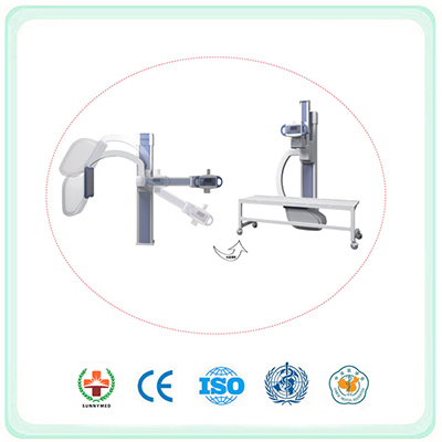
SDR01 U-arm CCD Detector Digital Radiography Equipment
Product Type: C-arm x-ray machine
Place of origin: China (Mainland) Guangdong
Model No:SDR01
Price Terms: FOB Guangzhou
Payment Terms: T/T.L/C.Western Union.MoneyGram
Package: Carton
Minimum Order: 1 set/sets
Delivery Time: 7-10 working days
Brand Name: SUNNY
Introduction:
UC-arm Digital Radiography System X-RAY detector uses KODAK VHD CCD by way of camera shot sensor, KODAK VHD CCD mainly uses in the outer space scientific research, has the superelevation sensitivity and extremely reliability, ultra long service life, This DR system CCD detector compares with ordinary CCD detector has a higher sensitivity and more superior image quality, compares the flat detector has the longer service life and a higher reliability, higher spatial resolution and extremely low use-cost.
Application:
Used to all over filming inspection: chest, skull, abdomen, spine, pelvis and limbs etc.
Parameter
Import CPI high frequency and high voltage generator 1 Power: 32KW;
Work frequency: 100KHz
Photographing (mAs): 500mAs
Photographing kV: 40~150kV
Import Toshiba 7242EX tube 1 Focus: 0.6/1.5mm
Photographing kv: 40~125kV
Heat Capacity: 200kHU
The up & down moving scope of the transverse arm:(65-165)cm±3cm ,continuously adjustable;
Transverse arm rotation around rotation center(rotate to the safety height):(0~105)°±2°;
Screen focus distance:(100-180)cm±5cm, continuously adjustable;
KODAK single CCD+ CsI
Image detector 1 Photographing size: ≥17*15 inch
Image pixels: ≥9 million
Limit space resolution: ≥3.4 Lp/mm
A/D transfer coefficient: 16bit
Specification:
1. Power-driven photographing shell 1 Sickle –Style arm photographing shelf.
2. Photographing bed 1 Photographing shelf and photographing bed: the moving up & down, left & right and rotation of the transverse arm should be steady.
3. Automatic exposure system 1 Import solid-state ionization chamber automatic exposure system
4. Clear cmos.
5. Automatic exposure.
6. Software 1 For real automatic exposure software system, doctors only need to press down hand brake to finish exposure operation instead of setting exposure parameter manually.
7. Image-collecting work station 1 Dell work station: AMD 2.5GHz, 1GB RAM, hard disk 320GB, 19 inch liquid crystal detector (1 set),100M network interface, DICOM3.0 interface.
8. Image collecting and disposing software 1 automatic /automatic window width / window level, preset window width / window level, positive and negative image turning, image turning, image rotation, image amplification and roaming, labeling and measuring, splitting, automatic electron cutting, part amplification, recovering original image.
9. Edge enhancement: automatically distinguish and analyze image, enhance image edge sharpness.
10. Dynamic scope optimization: automatically compress dynamic curve of original images for convenient diagnose.
11. High-frequency image enhancement:enhance image contrast,improve detail resolution, obviously improve small images such as bone trabecula's .
12. Noise inhibition: utomatically filter mixed signal, obviously reduce image noise,improve image signal-to-noise ratio.
13. Parts:
(1) 1 Import bucky; import beam limit device; high speed data transmission wire.
(2) Optional digital image work station software.
(3) K-PACS image storage transmission system.
(4) Film printer.
|


